The test kit utilize purified chlamydia trachomatis antibody antigen, based on gifa principle and use indirect method to detect urea plasma urealyticum antibody in human serum.
Test procedure
1. Remove the test kit and put it at room temperature (for about 20-30mins) before testing.
2. Add 1 drop of wash buffer into the hole and allow the liquid soak the membrane completely.
3. Add 150the serum specimen into the hole and allow it to sufficient absorption.
4. Add 3 drops of colloidal conjugate into the hole and allow it to sufficient absorption.
5. Add 3 drops of wash buffer into the hole and allow it to sufficient absorption. Read the results within 3 minutes.
Storage
The kit can be stored at room temperature. The test kit is stable through the expiration date (18 months) marked on the foil pouch. Do not freeze. Do not store the test kit in direct sunlight.
As for urine reagent analysis strip uncut sheets , we have various parameters ranging from 1 to 12 .
We can provide you strip and uncut sheet urs format with high quality and competitive price
Principle and expected values
Urs ascorbic acid: this test involves decolorization of tillmanreagent. The presence of ascorbic acid causes the color of the test field to change from blue-green to orange. Glucose: this test is based on the enzymatic reaction that occurs between glucose oxidase, peroxidase and chromogen. Glucose if first oxidized to produce gluconic acid and hydrogen peroxide in the presence of glucose oxidase. The hydrogen peroxide reacts with potassium iodide chromogen in the presence of peroxidase. The extent to which the chromogen is oxidized determines the color which is produced, ranging from green to brown. Low amounts of glucose are normally excreted in urine.3 glucose concentrations as low as 100 mg/dl, read at either 10 or 30 seconds, may be considered abnormal if results are consistent. At 10 seconds, results should be interpreted qualitatively. For semi-quantitative results, read at 30 seconds only.
Urs bilirubin: this test is based on azo-coupling reaction of bilirubin with diazotized dichloroaniline in a strongly acidic medium. Varying bilirubin levels will produce a pinkish-tan color proportional to its concentration in urine. In normal urine, no bilirubin is detectable by even the most sensitive methods. Even trace amounts of bilirubin require further investigation. Atypical results (colors different from the negative or positive color blocks shown on the color chart) may indicate that bilirubin-derived bile pigments are present in the urine specimen, and are possibly masking the bilirubin reaction.
Feline immunodeficiency virus ab test is a sandwich lateral flow immunochromatographic assay for the qualitative detection of feline immunodeficiency virus antibody (fiv ab) in catsblood.
Principle of the assay
Feline immunodeficiency virus ab test is based on sandwich lateral flow immunochromatographic assay. The test device has a testing window. The testing window has an invisible t (test) zone and c (control) zone. When sample is applied into the sample hole on the device, the liquid will laterally flow on the surface of the test strip. If there is enough fiv antibody in the sample, a visible t band will appear. The c band should always appear after a sample is applied, indicating a valid result. By this means, the device can accurately indicate the presence of fiv antibody in the sample.
Interpretation of results
Positive: the presence of both c band and t band, no matter t band is clear or vague.
Negative: only clear c band appears.
Invalid: no colored band appears in c zone, no matter whether t band appears.
Storage
The kit can be stored at room temperature (2-30â°c). The test kit is stable through the expiration date (18 months) marked on the foil pouch. Do not freeze. Do not store the test kit in direct sunlight.
The One Step TORCH IgG/IgM Test is a panel of rapid qualitative lateral flow test designed for the quantitive detection of IgG and IgM antibodies to Toxoplasma gondii (TOXO), Cytomegalovirus (CMV), Rubella, Herpes Simplex Virus (HSV) in human serum/plasma samples.
The amp gold rapid screen test is a qualitative competitive binding immunoassay for determination of amphetamine in urine.
Amp principle
The amp gold rapid screen test is a chromatographic absorbent device in which drug or drug metabolites in a sample compete with drug antigen immobilized on a porous membrane support for limited antibody sites. Labeled antibody-dye conjugate mixes with sample specimen and binds to the free drug presented in sample forming an antibody-antigen complex. This complex prevents the formation of pink color bands in the test zone immobilized antigen conjugate when the drug is presented in the sample urine above the detection levels (1000ng/ml for amphetamine). Unbound dye conjugate binds to the reagent in the control zone and produces a pink-rose color band, demonstrating that the reagents and device are functioning correctly.
Amp reagents and materials provided
1. Test device a pouched cassette contains a single test for amphetamine
2. Dropper a transfer pipette seal in foil pouch together with test device
3. Operating instructio materials required but not provided
Amp materials required but not provided
5348 Other Medical Equipments Suppliers
Short on time? Let Other Medical Equipments sellers contact you.
Home Menopause Test Strip is an in vitro diagnostic (IVD) qualitative test for rapid detection of human Follicle Stimulating Hormone (FSH) in urine at a sensitivity of 25mIU/ml.
SUMMARY AND EXPLANATION OF THE TEST
Follicle Stimulating Hormone (FSH) is a peptide hormone produced in the pituitary gland of the brain. It is normally present in the blood or urine varying in concentration with the stage of the menstrual cycle. When estrogen levels drop, FSH is released from the pituitary gland indicating that either a woman in mid-menstrual cycle or the onset of perimenopause. During early menopause, changes take place in the balance of hormones that regulate and control menstrual cycles. As a woman grows older and passes out of childbearing stage of life, the ovaries gradually make less of the hormone estrogen and FSH increases.FSH normally regulates the growth and development of an egg. Once this part of the monthly cycle is complete, FSH production is stopped and it returns to normal. As the body decreases estrogen production with age, more FSH is made. Over time these hormone changes cause menstrual periods to stop completely and "menopause" has occurred. The slow change in ovary function can happen between 2 and 10 years before the final period. This early stage before menopause is called perimenopause. During this stage, the levels of FSH may rise to positive levels and slowly return to normal, causing irregular or missed periods. The testing for FSH should be performed twice to help identify the levels of FSH throughout a menstrual cycle.
PRECAUTION
1. For in vitro diagnostic use only.
2. Do not use after expiration date.
3. Test device should remain sealed until ready for use. Do not use if pouch is damaged or opened.
4. Read this instruction carefully before performing the test.
For the qualitative assessment of hbsab in humanserum, plasma or whole blood
Intended use
Rapid hbsab test is a chromatographic immunoassay for qualitative detection of the surface antibody of hepatitis b virus (anti-hbs) in human whole blood samples. It is intended for use in medical institution as an aid for diagnosis and management of patients related to infection with hepatitis b as well for screening of blood donors or blood products
Principle
Rapid hbsab test employs chromatographic lateral flow device. Colloidal gold conjugated surface antigen reactive to anti-hbs (sag-au) are dry-immobilized onto a nitrocellulose membrane strip. When the sample is added, it migrates by capillary diffusion through the strip rehydrating the gold conjugate. If present, anti-hbs will bind with the gold conjugated antigens forming particles. These particles will continue to migrate along the strip until the test zone (t) where they are captured by hbsag immobilized there and a visible red line appears. If there is no anti-hbs in sample, no red line will appear in the test zone (t). The gold conjugate will continue to migrate alone until is captured in the control zone (c) from immobilized goat, anti-hbs antibody and aggregating in a red line, which indicates the validity of the test
Storage
Store the test device at 4 to 30â°c. Do not freeze.
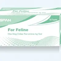
Feline panleucopenia virus ag test is a sandwich lateral flow immunochromatographic assay for the qualitative detection of feline panleucopenia virus (fpv ag) in cats feces or vomit.
Principle of the assay
Feline panleucopenia virus(fpv) ag test is based on sandwich lateral flow immunochromatographic assay. The test device has a testing window. The testing window has an invisible t (test) zone and c (control) zone. When sample is applied into the sample hole on the device, the liquid will laterally flow on the surface of the test strip. If there is enough panleucopenia virus antigen in the sample, a visible t band will appear. The c band should always appear after a sample is applied, indicating a valid result. By this means, the device can accurately indicate the presence of panleucopenia virus antigen in the sample.
Interpretation of results
Positive: the presence of both c band and t band, no matter t band is clear or vague.
Negative: only clear c band appears.
Invalid: no colored band appears in c zone, no matter whether t band appears.
Storage
The kit can be stored at room temperature (2-30â°c). The test kit is stable through the expiration date (18 months) marked on the foil pouch. Do not freeze. Do not store the test kit in direct sunlight.
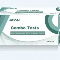
Fiv ab + felv ag combined rapid test is a combined cassette to differentially diagnose the presence of feline immunodeficiency antibody and feline leukemia virus antigen in cats blood.
Assay time: 10-15 min
Sample: serum, plasma or whole blood
2. Principle of the assay
Fiv ab + felv ag combined rapid test is based on sandwich lateral flow immunochromatographic assay. The test device has two testing windows. Each testing window has an invisible t (test) zone and c (control) zone. When sample is applied into the sample hole on the device, the liquid will laterally flow on the surface of the test strip. If there is enough fiv antibody or felv antigen in the sample, a visible t band will appear in the corresponding testing window. The c band should always appear after a sample is applied, indicating a valid result. By this means, the device can accurately indicate the presence of fiv antibody or felv antigen in the sample.
3. Kit component
- 10ã?foil pouches, each containing a cassette,pipette and desiccant
The hcv gold rapid screen test (rst) is a chromatographic immunoassay (cia) for direct qualitative detection of antibodies to
Hepatitis c type virus (hcv) in human serum/ plasma and whole blood.
Principle
Hcv rst is a chromatographic immunoassay (cia) for the detection of antibodies to hcv in human serum/plasma and whole blood. Hcv recombinant antigens are precoated onto membrane as a capture reagent on the test region. During the test, specimen is allowed to react with the colloidal gold particles, which have been labeled with hcv recombinant antigens. If antibodies to hcv present, a pink colored band will develop on the membrane in proportion to the amount of hcv antibodies present in the specimen .Absence of this pink colored band in the test region suggests a negative result. To serve as a procedural control, a purple colored band in the control region will always appear regardless the presence of antibodies to hcv.
Reagents and materials provided
1. One pouched cassette with desiccant.
2. Blood diluent in a dropper bottle, stored at 4-30â°c.
3. One piece of operating instruction.
Warning and precautions
1. For in vitro diagnostic uses only.
2. All patient samples should be treated as if capable of transmitting diseases.
3. Do not interchange reagents from different lots or use test kit beyond expiration date.
4. Icteric, lipemic, hemolysed, heat treated and contaminated sera may cause erroneous results.
Storage
The kits should be stored at temperature 4-30â°c, the sealed pouch for the duration of the shelf life (24 months).
The hcv gold rapid screen test (rst) is a chromatographic immunoassay (cia) for direct qualitative detection of antibodies to
Hepatitis c type virus (hcv) in human serum/ plasma and whole blood.
Principle
Hcv rst is a chromatographic immunoassay (cia) for the detection of antibodies to hcv in human serum/plasma and whole blood. Hcv recombinant antigens are precoated onto membrane as a capture reagent on the test region. During the test, specimen is allowed to react with the colloidal gold particles, which have been labeled with hcv recombinant antigens. If antibodies to hcv present, a pink colored band will develop on the membrane in proportion to the amount of hcv antibodies present in the specimen .Absence of this pink colored band in the test region suggests a negative result. To serve as a procedural control, a purple colored band in the control region will always appear regardless the presence of antibodies to hcv.
Reagents and materials provided
1. One pouched cassette with desiccant.
2. Blood diluent in a dropper bottle, stored at 4-30â°c.
3. One piece of operating instruction.
Warning and precautions
1. For in vitro diagnostic uses only.
2. All patient samples should be treated as if capable of transmitting diseases.
3. Do not interchange reagents from different lots or use test kit beyond expiration date.
4. Icteric, lipemic, hemolysed, heat treated and contaminated sera may cause erroneous results.
Storage
The kits should be stored at temperature 4-30â°c, the sealed pouch for the duration of the shelf life (24 months).
The hcv gold rapid screen test (rst) is a chromatographic immunoassay (cia) for direct qualitative detection of antibodies to
Hepatitis c type virus (hcv) in human serum/ plasma and whole blood.
Principle
Hcv rst is a chromatographic immunoassay (cia) for the detection of antibodies to hcv in human serum/plasma and whole blood. Hcv recombinant antigens are precoated onto membrane as a capture reagent on the test region. During the test, specimen is allowed to react with the colloidal gold particles, which have been labeled with hcv recombinant antigens. If antibodies to hcv present, a pink colored band will develop on the membrane in proportion to the amount of hcv antibodies present in the specimen .Absence of this pink colored band in the test region suggests a negative result. To serve as a procedural control, a purple colored band in the control region will always appear regardless the presence of antibodies to hcv.
Reagents and materials provided
1. One pouched cassette with desiccant.
2. Blood diluent in a dropper bottle, stored at 4-30â°c.
3. One piece of operating instruction.
Warning and precautions
1. For in vitro diagnostic uses only.
2. All patient samples should be treated as if capable of transmitting diseases.
3. Do not interchange reagents from different lots or use test kit beyond expiration date.
4. Icteric, lipemic, hemolysed, heat treated and contaminated sera may cause erroneous results.
Storage
The kits should be stored at temperature 4-30â°c, the sealed pouch for the duration of the shelf life (24 months).
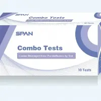
Cpv + ccv ag combined test is a combined cassette to differentially diagnose the presence of canine parvovirus antigen and canine coronavirus antigen in dogs feces or vomit.
Assay time: 5 -10 min
Sample: feces or vomit
2. Principle of the assay
Cpv + ccv ag combined test is based on sandwich lateral flow immunochromatographic assay. The test device has two testing windows. Each testing window has an invisible t (test) zone and c (control) zone. When sample is applied into the sample hole on the device, the liquid will laterally flow on the surface of the test strip. If there is enough cpv antigen or ccv antigen in the sample, a visible t band will appear in the corresponding testing window. The c band should always appear after a sample is applied, indicating a valid result. By this means, the device can accurately indicate the presence of cpv antigen or ccv antigen in the sample.
3. Kit component
- 10ã?foil pouches, each containing acassette, pipette and desiccant
Cpv + ccv +giardia ag triple test is a combined cassette to differentially diagnose the presence of canine parvovirus antigen, canine coronavirus antigen and giardia antigen in dogs feces or vomit.
Assay time: 5-10 min
Sample: feces or vomit
2. Principle of the assay
Cpv + ccv + giardia ag triple test is based on sandwich lateral flow immunochromatographic assay. The test device has three testing windows. Each testing window has an invisible t (test) zone and c (control) zone. When sample is applied into the sample hole on the device, the liquid will laterally flow on the surface of the test strip. If there is enough cpv antigen, ccv antigen or giardia antigen in the sample, a visible t band will appear in the corresponding testing window. The c band should always appear after a sample is applied, indicating a valid result. By this means, the device can accurately indicate the presence of cpv, ccv or giardia antigen in the sample.
3. Kit component
- 10ã?foil pouches, each containing onecassette and a desiccant
Cdv + cpv ag combined rapid test is a combined cassette to diagnose the presence of canine disteimper virus antigen and canine parvovirus antigen in dogs secretions, serum or feces.
Assay time: 5 - 10 min
Sample: secretions, serum or feces
2. Principle of the assay
Cdv + cpv ag combined rapid test is based on sandwich lateral flow immunochromatographic assay. The test device has two testing windows. Each testing window has an invisible t (test) zone and c (control) zone. When sample is applied into the sample hole on the device, the liquid will laterally flow on the surface of the test strip. If there is enough cdv antigen or cpv antigen in the sample, a visible t band will appear in the corresponding testing window. The c band should always appear after a sample is applied, indicating a valid result. By this means, the device can accurately indicate the presence of cdv antigen or cpv antigen in the sample.
3. Kit component
- 10ã?foil pouches each containing a cassette, pipette and desiccant
The one step toxo igg/igm test is a rapid chromatographic immunoassay for the qualitative detection of antibodies (igg and igm) to toxoplasma gondii in whole blood /serum / plasma to aid in the diagnosis of toxo infection. Toxoplasma gondii is an obligate intracellular protozoan parasite with a worldwide distribution. Serological data indicates that approximately 30% of the population of most industrialized nations is chronically infected with the organism. A variety of serologic tests for antibodies to toxoplasma gondii have been used as an aid in diagnosis of acute infection and to assess previous exposure to the organism. These tests are the sabin-feldman dye test, direct agglutination, indirect hemagglutination, latex agglutination, indirect immunofluorescence, and elisa. Recently, lateral flow chromatographic immunoassay, such as the toxo igm/igg rapid test (whole blood/serum/plasma) was introduced into the clinic for the serodiagnosis of toxoplasma gondii infection. One step toxo igg/igm test is a simple, visual qualitative test that detects toxo antibodies in human whole blood/serum/plasma. The test is based on immunochromatography and can give a result within 15 minutes.
Storage and stability
Store as packaged in the sealed pouch at room temperature or refrigerated (4-30â?? or 40-86â??). The test device is stable through the expiration date printed on the sealed pouch.
The test must remain in the sealed pouch until use.
For the qualitative assessment of hbsag in human serum, plasma or whole blood
Intended use
The hbsag rapid test is a chromatographic immunoassay (cia) for direct qualitative detection of hepatitis b type virus surface antigen (hbsag) in human serum /plasma and whole blood.
Principle
The hbsag rst is a chromatographic immunoassay (cia) for the detection of surface antigens of hepatitis b in human serum/plasma and whole blood. Specific antibody against hbsag is pre-coated onto membrane as a capture reagent on the test region. During the test, specimen is allowed to react with the colloidal gold particles, which have been labeled with anotherr specific antibody. If hbsag is present, a pink colored band will develop on the membrane in proportion to the amount of hbsag presented in the specimen. Absence of this pink colored band in the test region suggests a negative result. To serve as a procedural control, a pink colored band in the control region will always appear regardless the presence or absence of hbsag.
Reagents and materials provided
1.One sealed pouched cassette with desiccant and a disposable pipette..
2.Blood diluent in a dropper bottle. Store at 2-8â°c.
3.One piece of operating instruction with 40 test pouches..
The leishmania igg/igm rapid test is a lateral flow immunoassay for the qualitative detection of antibodies including igg and igm to the subspecies of the leishmania donovani (l. Donovani), the visceral leishmaniasis causative protozoans in human serum or plasma. This test is intended to be used as a screening test and as an aid in the diagnosis of the disease of visceral leishmaniasis. Any reactive specimen with the leishmania igg/igm rapid test must be confirmed with alternative testing method(s).
Summary and explanation of the test
Visceral leishmaniasis, or kala-azar, is a disseminated infection caused by several subspecies of the l. Donovani. The disease is estimated by the world health organization (who) to affect approximately 12 million people in 88 countries1. It is transmitted to humans by bites of the phlebotomus sandflies, which acquire infection from feeding on infected animals. Though it is a disease for poor countries, in southern europe, it has become the leading opportunistic infection in aids patients2-3.
Identification of l. Donovani organism from the blood, bone marrow, liver, lymph nodes or the spleen provides a definite means of diagnosis. However, these test methods are limited by the sampling method and the special instrument requirement. Serological detection of anti-l. Donovani ab is found to be an excellent marker for the infection of visceral leishmaniasis. Tests used in clinic include: elisa, fluorescent antibody and direct agglutination tests4-5. Recently, utilization of l. Donovani specific protein in the test has improved the sensitivity and specificity dramatically6-7.
The leishmania igg/igm rapid test is a recombinant protein based serological test, which detects antibodies including igg, igm and iga to the l. Donovani. This test provides a reliable result within 10 minutes without any instrumentation requirements.
One step h. Pylori ab wb /serum/plasma test is a rapid test for the qualitative detection of antibodies specific to helicobacter pylori in human serum/plasma and whole blood. In this test kit, the h. Pylori antigencolloid gold conjugate and specimen moves along the membrane chromatographically to the test region and forms a visible line as the antigenantibodyantigen gold particle complex forms with high degree of sensitivity and specificity. This one step test only takes about 1015 minutes. Test results are read visually without any instrumentation.
Specimen collection & preparation
Both whole blood (with or without anticoagulant), serum and plasma specimens can be used with this assay. Simply follow the standard clinical produres to collect whole blood, serum or plasma specimens. If the specimen cannot be tested on the day of collection, store the serum/plasma specimen in a temperature of 28 âºc for up to 72 hours. Stir and bring the specimens to room temperature before testing. Do not freeze whole blood specimens. Do not freeze and thaw the specimen repeatedly. Attention: specimens and all materials coming in contact with them should be handling and disposed of as if capable of transmitting infection. Avoid contact with skin by wearing gloves and proper laboratory attire.
The hcg s/u gold rapid screen test(rst)is a chromatographic immunoassay for the early detection of human chronic gonadotropin (hcg) in serum/plasma or urine specimens.
Introduction
Hcg is a glycoprotein hormone secreted by the developing placenta during pregnancy. The concentration of hcg in serum is approximately equal to the concentration in urine. The concentrations of hcg in urine and serum continue to rise during the first trimester of pregnancy to as high as 100,000 miu/ml. Hcg appears in urine shortly after conception, and continues to increase during the early stages of pregnancy, making it an excellent indicator for the detection of pregnancy.
Principle
The membrane of the test device was coated with anti hcg antibodies on the test region and goat anti mouse igg antibodies on the control region. During the test, urine specimen is allowed to react with the hcg monoclonal antibody-colloid gold conjugate, which was pre-dried on the test strip. The mixture then moves upward on the membrane chromatographically by capillary action. For a positive specimen, the conjugate binds to the hcg forming an antibody-antigen complex. This complex is captured by anti hcg antibody immobilized on the test region (t) and produces a pink color band when hcg concentration is equal to or greater than 25miu/ml. Absence of this colored band in the test region suggests a negative result. To serve as a procedural control, a colored band at the control region(c) will always appear regardless the presence or absence of hcg.
Storage
The kits should be stored at temperature 4-30â°cï¼? the sealed pouch for the duration of the shelf life (24months).
5348 Other Medical Equipments Suppliers
Short on time? Let Other Medical Equipments sellers contact you.